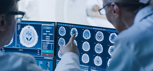
Specialist - Diagnostic Radiology
Back to School
Parents should encourage their children to adjust to an earlier bedtime and earlier waking habits at least a week before school starts.
Overview
Radiology, also called diagnosis imaging, is a medical discipline that uses imaging technology to diagnose and treat certain diseases in humans. It captures images of the affected body parts through various imaging technologies like X-ray, Ultrasound, CT (computed tomography), etc. Doctors and healthcare professionals who perform radiology are known as Radiologists. The medical procedure of Radiology can be categorized into two broad categories. We'll discuss them one by one in this article.
Diagnostic Radiology
It enables healthcare professionals to detect the structure inside the body and hence any injury or disease. Specialized doctors, also called Diagnostic Radiologists, comprehend these images and take further required actions. These allow the doctors to diagnose the causes of symptoms occurring to the patient. They can also watch if the patient's body responds positively to the given treatment of the disease and let them screen for other serious illnesses. The images are taken through various methods like:
1. X-rays: X-rays are done by transmitting X-rays through the patient's body onto a detecting device. The image of the concerned body part is obtained by the nature of the rays that got absorbed or scattered and hence remained undetected.
2. Computed Tomography (CT): CT imaging combines X-rays with a computing algorithm to take the image of the body part. CT can detect more amplified variations of the disease.
3. MRI (Magnetic Resonance Imaging) and MRA (Magnetic Resonance Angiography): This involves use of strong magnetic or radio waves onto the body tissue of the patient. These scans provide the best results for detecting any disease in the soft tissues. These don't utilize ionizing radiations like that of an X-ray.
4. Fluoroscopy: Fluoroscopy is a particular application of regular X-rays. The X-rays are passed through the patient's body, and multiple images are obtained on the screen to produce real-time moving objects inside the body.
5. Ultrasound: This uses high-frequency sound waves, also called ultrasound, to see real-time soft tissue structure in the body and obtain its image. This process requires the patient to lie down as the healthcare professional uses a hand probe called a transducer that is moved along the affected area.
Interventional Radiology
Here, a minimal insertion is performed by the doctor in order to obtain the image of the affected areas. Specialized catheters, wires, and various other smaller tools are used for this purpose and imaging technologies like CT, ultrasound, and MRI are applied to direct the process.
• Needle biopsy of specific organs like thyroid glands.
• Feeding tube placement
• Breast biopsy, guided by ultrasound techniques
Services Offered